LABORATORY: INFN CHNet
NAME OF THE INSTRUMENT
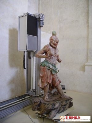
Instrument for in-situ computed tomography, developed by the CHNet laboratories.
GENERAL DESCRIPTION
X-ray Computed Axial Tomography (XCT) is a non-destructive diagnostic technique capable of visualizing the internal structure of the analyzed objects in 3D. The CT scan is typically performed on movable goods (very rarely and with ad hoc designed systems, equipped with an X-ray source with energy of some MeV, it can be performed on architectural elements such as poles or columns, or large statues, but only if around there is enough space for mounting the system and if the thicknesses and materials allow it).
The XCT is suitable for a great variety of artifacts and materials, but it is difficult to carry out analyses on all types of objects with the same system. The major limitations are given by the density of the material (metals), by the average total thickness (solid marble), by the size (large objects).
The systems developed in the CHNet laboratories are suitable for medium-light materials such as wood, clay, terracotta etc ... and make it possible to carry out analyses on both statues and paintings on wood. XCT measures the local density of the material but has a limited ability to distinguish between materials. The materials with sufficiently different densities clearly differ from each other: light metals from heavy metals, wood from metal, solid cavities, plaster on wood, etc. Two portable systems have been developed, one for small-medium sized objects, the other for large objects.
TECHNICAL DESCRIPTION
High resolution transportable system for medium sized objects
- Detector mounted on a translation system consisting of 2 orthogonal axes with range of 30 cm
- Detector: flat panel VARIAN PS2520D
- FOV: 25 x 20 cm2
- Pixel size: 127 μm
Mobile system for RX / CT of large objects
- System with two orthogonal axes, each with range of 1.5 m, on which a flat-panel is mounted.
- The system can be transported on site by means of a van with a load compartment of at least 2 m deep.
- The source usually used is a Gilardoni RX tube with a large emission cone with a maximum voltage of 200 kVp.
- Detector: Flat-panel Hamamatsu C10900D
- FOV: 12 x 12 cm2
- Pixel size: 100 μm
Referents:
Lisa Castelli lisa.castelli@fi.infn.it
Francesco Taccetti ftaccetti@fi.infn.it
