LABORATORIO: CNR ISPC – XRAYLab
NAME OF THE INSTRUMENT
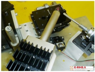
LE-XRF spectrometer developed at XRAYLab of CNR-ISPC
GENERAL DESCRIPTION
Compositional studies applied to the surface of objects of archeological or historical and artistic value provides important insight into the nature of the materials used and the fabrication process. Typically the instrumentation used for such analysis are located within scientific laboratories (as is the case for the PIXE methodology that requires synchrotron radiation) and cannot be displaced. The XRF method presented here is particularly well suited for the investigation of the first surface layers. As well known, conventional XRF is not surface sensitive and probes up to several tens of microns within the samples. Additionally, even though XRF is generally a very efficient technique, the detection limits in the lower energy region of its spectra are often so high that light elements (like Na and Mg) cannot be detected at all. These limitations arise both from the presence of a high background due to diffusion of the higher part of the primary beam spectrum, and absorption in the air, if it is present in the detection path.
XRAYlab of ISPC developed a mobile XRF spectrometer particularly suited for the compositional analysis of surfaces. It employs a low energy primary beam (operating at 8KV and 2mA) of probing depth limited to the first few microns for heavy matrix samples (such as metals) and lower than 20 microns even for light matrices (like glasses or ceramics). Furthermore, since the primary beam only contains Xrays of energy than 8KeV, the low energy side of the spectrum is affected by less noise and lighter elements are more effectively revealed (starting from Na, near 1keV). Blowing helium between the sample and the detector further increase the detection efficiency of light elements.
TECHNICAL DESCRIPTION
The LE_XRF portable system developed within the XRAYlab of ISPC exploits an X-ray tube operating at low voltage (8KV) and high current (2mA). The detection of X-ray fluorescence, emitted from the sample after interaction with the primary beam, is performed by an SDD detector with active area of 80mm2 . The beam spot on the measuring sample has diameter of around 3mm. To minimized absorption by air, a flux of helium is blown in the detection path during measurements, allowing detection of low energy emission lines from lighter elements (starting from sodium at ~1KeV). Elements are identified from their K-emission (low to medium atomic numbers, such as Na, Al, P, Ca, Ti, Fe, Mn), their M-emission (heavy elements like Hg, Pb and Au) or their L-lines (for medium-heavy elements like Ag and Sn).
Referent:
Paolo Romano francescopaolo.romano@cnr.it
