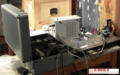
LABORATORY: CNR INO
NAME OF THE INSTRUMENT
FD-OCT instrument; TD-OCT instrument
GENERAL DESCRIPTION
Optical Coherent Tomography (OCT), typically applied in the biomedical field, is an interferometric technique that provides high-resolution stratigraphic sections of diffusing or semi-transparent objects. The spectral bandwidth of the source, emitting either in the visible or in the near infrared region, sets the in-depth resolution. OCT has found valuable application in painting diagnostics, allowing for the stratigraphy of pictorial layers in a completely non-invasive way. In-depth analysis is particularly useful for measuring and documenting micrometric variations in layers’ thickness during cleaning intervention, which involves the selective removal of deteriorated patinas or deposits (paints, oxalates, overpainting) from the surface. However, the visualization of the stratigraphy by OCT can be hindered by the presence of highly scattering interfaces, which prevent the propagation of the incident radiation in depth. To overcome this limit, the OCT set-up was coupled to a confocal scanning design, allowing to focus the incident beam inside the sample on the interface of interest, maximizing, thus, the back-scattered signal.
TECHNICAL DESCRIPTION
The penetration depth of an optical probe depends both on the properties of the analysed material (chemical composition, thickness, ...) and on the wavelength of the radiation. In this regard, CNR-INO provides two OCT devices, operating at 1300 nm (frequency domain, FD-OCT) and 1550 nm (confocal time-domain TD-OCT). The instruments’ output is a set of in-depth images of the sample (also termed stratigraphic or tomographic images) constituting the so-called tumocube, which can be analysed according to any section (xz, yz, xyThe FD-OCT device allows acquiring volumes with 10 × 10 × 3.5 mm3 maximum size, with lateral and axial (in depth) resolution of 13 microns and 5.5 microns in air, respectively. The TD-OCT instrument provides volumes with 25 × 25 × 1 mm3 maximum size, with lateral and axial resolution of about 2.5 microns and 10 microns in air, respectively. For axial measurement, the measured optical distance has to be divided by the refractive index of the analysed material to obtain the real thickness.
Depending on the characteristics of the artwork, one or the other of the two OCT system is used.
Referent:
Raffaella Fontana raffaella.fontana@ino.cnr.it
