LABORATORY: CNR ISPC
NAME OF THE INSTRUMENT
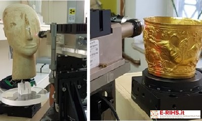
Mobile MA-XRF rotational scanner developed within the XRAYLab of CNR-ISPC
GENERAL DESCRIPTION
Macro X-ray Fluorescence (MA-XRF) is a non-destructive analytical method based on the scanning of the whole sample surface through an X-ray beam of size in the hundreds of micron range. The sample's X-ray fluorescence radiation, induced by the primary beam, is recorded as a function of the position, allowing the imagining of the detected (inorganic) chemical elements and their spatial distribution on the sample's surface. Such information is of remarkable interest in the study of artworks and archeometric materials.
Up until now, the MA-XRF method has been applied only to flat surfaces (such as paintings, typically), however many objects with complex geometry would also benefit from the investigation with this technique. . Combining rotational MA-XRF imaging with high resolution photogrammetry method results in the reconstruction of the full 3D model of the measured object. MA-XRF and photogrammetry data are processed together by a specifically tailored algorithm so to output a virtual reconstruction of the elemental maps projected directly on the studied object. During the measurement, to avoid accidental contacts, various dynamical correction system are in operation to maintain a constant distance between the spectroscopic head and the studied part of the object.
Security stops are automatically enforced as an extra precaution in case this distance is reduced below the confidence limit. The spectral distribution MA-XRF images are available in real time to the user already during the measurements. All materials of inorganic nature can be analyzed. Optimal cases of application are pictorial works on any three-dimensional support.
TECHNICAL DESCRIPTION
The mobile MA-XRF rotational scanner developed within the XRAYLab of CNR-ISPC is a mechatronic system with real-time technology. The system comprises a spectroscopic head with a microfocus Rh anode X-ray tube of 30W power coupled with polycapillary focalizing optics. A second X-ray tube with a Cr anode can be switched in place of the Rh one for higher resolution in the acquisition of the distribution of low atomic number chemical elements, with characteristic lines in the low energy range. Furthermore, the combined use of the two X-ray source allows to discriminate the distribution of the same element across different depth layers of the object, as the two sources have different penetration depth. A typical example is the distinction between lead present in the preparation base layer versus that used for shading contrasts. The X-ray fluorescence induced by the primary beam is detected in event mode by a multi-detector system operating in parallel, that includes up to two SDD of 50 mm2 active area and energy resolution <130 meV at 5.9 KeV. The spectroscopic head is mounted on a 3-axis system (XYZ) with span of 110x70x20 cm3 that allows to operate the scan in continuous mode aT high speed (up to 100mm/sec) with residence time of up to 10 msec per pixel. The measured object is positioned on a rotational stage with 360deg span and maximal resolution of 1 mdeg.
The system is equipped with a central CPU for the control of the measure operative parameters and security systems. The CPU manages the scanning sequence; automatically regulates the parameters of the source and detectors to avoid degradation of both the dead time and energy resolution of the recorded XRF spectra of each pixel; manages a 750 frames/sec laser interferometer for the dynamical correction of the distance between the object and the spectroscopic head so to avoid accidental contact; saves the scan coordinates to allow resuming the analysis of the same position in case of prolonged pauses or system arrest (such as overnight interruptions). Moreover, the CPU performs dynamical spectral analysis pixel per pixel by applying an accurate fitting procedure that outputs elemental images in real time, without artifacts. The scanning system is also equipped of a real-time image analysis software that can: correlate images of different elements (RGB correlations), apply logical-mathematical operators, statistical analysis (such as PCA, ICA, NNMF), generate scatter plot and export single XRF spectra from a selected image area (ROI).
Referent:
Paolo Romano francescopaolo.romano@cnr.it
