LABORATORY: INFN CHNet
NAME OF THE INSTRUMENT
Scanner for in-situ digital radiography, developed by the CHNet laboratories
GENERAL DESCRIPTION
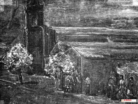
The X-ray radiography, normally applied to paintings on canvas and wood, provides information on the state of conservation of the artwork and can reveal important details such as the presence of underlying paintings, pentimenti and/or restoration interventions carried out in the past. In addition, the use of digital detectors allows not only to obtain an immediate result, but also to maintain a wide range of gray levels and to make the stitching of the radiographic images via software.
The scanner for in-situ radiography was designed and built to meet the need not to move the works from the place where they are stored, while having no limits in the size of the works to be analyzed.
TECHNICAL DESCRIPTION
The scanner for in-situ radiography consists of two independent aluminium units (each weighing less than 55 kg), one for handling the X-ray tube and the other for the digital detector. Typically, the two units are placed at 1 m from each other. The acquisition of the radiographies covers an area of 1 x 1 m2, however the scanning of larger paintings is possible. The scan is adapted based on the size of the painting, and the images obtained are automatically stitched at the end of the measurement. For example, scanning an area of 1 x 1 m2 requires 144 shots and about 3 hours.
The X-ray tube is a YXLON EVO 160D, maximum anode voltage 160 kV, maximum current 7 mA, air-cooled. The detector is a Teledyne DALSA RedEye200, consisting of a photodiode array with CMOS technology combined with a Gd2O2scintillator screen. It is made up of 1024 x 1000 pixels of 96 µm per side and the digitization depth is 12 bits/pixel.
Referents:
Lisa Castelli lisa.castelli@fi.infn.it
Francesco Taccetti ftaccetti@fi.infn.it

LABORATORY: CNR-ISPC – XRAYLab
NAME OF THE INSTRUMENT
X-Ray Digital Radiography developed at XRAYLab of CNR-ISPC
GENERAL DESCRIPTION
Digital radiography (DR) is an advanced imaging technique widely used within museums, with prime application on canvas and panel paintings. DR is particularly useful to qualitatively identify the materials, evaluate the state of preservation, to learn about the creative process of the artists and the techniques they used. Given its no-contact and not invasive nature, DR is exceptionally suited to investigate fragile and delicate works, such are for example paintings.
To obtain the radiography image the sample is illuminated by a not -collimated X-Ray beam, produced by a medium-power laboratory tube, so that the irradiated spot has linear dimensions of several centimeters. The incident beam is absorbed differently by the different layers of the sample, according to their thickness and density, thus producing a grey-scale image on an appropriate sensor situated behind the sample. The employment of detectors with wide active area (digital flat-panels of roughly 45x35 cm2 active area in the system of ISPC-CT) allows the acquisition of the radiography of a wide area of the sample in a single take and in reduced times (few tens of a second). In the case of large-scale artworks, several frames can be recorded in a mosaic-like pattern and the full image is later reconstructed by an automatic stitching software.
Synthetic guide for choosing the X-Ray Digital Radiography method of ISPC
Materials: inorganic materials with a high degree of opacity.
Optimal application: paintings on any support (except frescos), including large sizes (in stitching mode), (quasi) planar works in metal or wood.
Sample positioning: vertical.
Type of analysis: non-destructive and in-situ
Measuring times: variable, depending on sample size. Typically, less than a minute per single take (area of 43x35cm22 or 46x38 cm22 according to the detector used).
Active signal resolution: 2816 x 2304 pixel Pixel Matrix: 2816x2304 or 2560x3072 pixels. Pixel size: 154 or 140 microns.
Characteristics and parameters of the X source: Teledyne CPD160 tube, 160Kv e 6mA, Be or Al filters with different form factors. Beam spot: a few centimeters.
Detection system: (a) Teledyne GO-SCAN 4335, aSi flat-panel, 43x35cm2v2816x2304 pixels, pixel size 154 microns or (b) VIVIX-S 1417N, aSi flat-panel, 2560x3072 pixels, pixel size 140 microns.
Gray-scale depth: 16 bit.
Data interface: GigaE or WI-FI
Frame rate: typically, 0.3 sec.
Security: as per usual for all X-ray based methods. The measurements take place following the prescriptions of the qualified radioprotection expert, in appropriate premises with thick walls.
TECHNICAL DESCRIPTION
The DR (digital radiography) system of the laboratory XRAYLab of CNR-ISPC comprises an X-ray source with maximum operating parameters of 160kV and 6 mA, equipped with filters of different shapes (circular and rectangular) in either Be or Al. The exit beam has angles of 60° and 40° in the Y e X directions, respectively.
The radiography of works of larger sizes is acquired in successive frames, with the aid of an Al frame of 200x180cm2 placed behind the painting, and the resulting mosaic is then assembled by a stitching software at the end of the measurements.
The two flat-panel detectors can operate with Giga-Ethernet or wi-fi interface and are equipped with a battery of up to 8 hours autonomy (which enables operation even in those cases where a direct connection to the electrical network is not available). The measurements take place with the sample positioned between the X-Ray tube and the detector, with typical distance between the source exit and the screen in the order of 1.0-1.5 meters. In the case of paintings on canvas, the X-Ray tube is routinely operated at 30kV and 6mA, with exposition times between 30 and 60 seconds. The frame rate of the available detectors is in the range of 0.3 seconds, and their grey-scale dynamic range is of 16 bits. The whole system is operated remotely through a laptop.
Referent:
Francesco Paolo Romano francescopaolo.romano@cnr.it
